
ANZSCDB Image Award
Jump to: Instructions to Applicants | Award Winners

Instructions to Applicants
Purpose:
The aim of these awards is to showcase the beauty of Cell and Developmental Biology by celebrating the awe-inspiring imaging of our ANZSCDB members. Up to two awards will be available to recognise outstanding images or movies (i.e. live imaging) in Cell and Developmental Biology. We encourage submissions from all ANZSCDB members across Australia and New Zealand.
Eligibility:
· The applicant must be a financial member of ANZSCDB at the time of the application.
· Images must be taken using an optical or electron microscope. Macro photography is not eligible.
· Each applicant may submit a maximum of ONE image and ONE movie.
· No submission can receive the award more than once, although applicants can submit the same image/movie in different years.
· Each entry must be the original work of the applicant and must not contain any material that infringes copyright, trademark, privacy or other intellectual property rights.
· The applicant grants ANZSCDB the right to reproduce, publish, transmit or otherwise communicate to the public their entry, in whole or in part, in or using any media for any purpose without permission or payment.
Selection criteria:
The awards are based on the best image or movie in the fields of Cell Biology and/or Developmental Biology.
ANZSCDB is committed to the principles of fairness, transparency, equity and diversity, including gender equality, in assessing and administering this award.
Required documentation:
1. A copy of the image/movie file (format should be TIFF, PDF or video format. Images should be high resolution with a recommended file size ~3-4MB, movies no larger than 100MB, as .avi or .mp4). For large files, please provide a URL link to a shared folder for download (e.g. CloudStor, Google Drive). Other sharing services that require the receiver to create an account will not be accepted.
2. A brief title and description of the work (~100 words- detailing the microscope/technique used, sample/staining preparation etc).
Applications are to be submitted to: anzscdb@asnevents.net.au
Application closing date: Friday June 26th, 2026.
Judging:
Evaluation, shortlisting, and ranking of submissions will involve members of the ANZSCDB Committee and the President.
Entries will be assessed based on the overall aesthetics of the images/movie, technical difficulty in obtaining the image/movie, research relevance and creativity/originality.
In situations where there is a tied vote or a lack of consensus, the President may cast the deciding vote or may choose to extend each award to more than one candidate.
ANZSCDB reserves the right to award joint candidates, or not confer an award, in any given round.
Prize:
Winners will receive a certificate and a $250 cash prize. Winning Images/Movies will be highlighted in ANZSCDB promotional materials and websites. Please ensure that images/movies submitted will not infringe copyright or ownership rules if displayed or disseminated by the society (this may entail altering the content, e.g. colours or field of view, for published material).
2025 ANZSCDB Image Awards

“Branching out: A network of astrocytes and neurons” by Adelene Chiam, Stem Cell Models Group, College of Health and Medicine Wicking Dementia Research and Education CentrE, University of Tasmania
2D in vitro model of human iPSC-derived astrocytes and cortical neurons co-cultured in a 2:1 ratio. By DIV 21, neurons exhibited extensive neurite arborisation and strongly expressed the neurofilament heavy chain (NfH, magenta) which is a marker of neuron maturity. Importantly, astrocytes in co-culture adopted a highly stellate morphology reminiscent of that in vivo, as opposed to astrocytes in monoculture which typically appear polygonal. The astrocytes were also found to be predominantly dual-positive for the canonical astrocyte markers GFAP (red) and S100β (green). Fixed with 4% paraformaldehyde and imaged via immunofluorescence microscopy using Celldiscoverer7 (Zeiss).
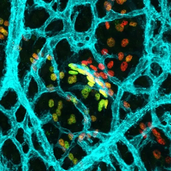
“Transcriptional Flow” by Genevieve Secker. University of South Australia and SA Pathology
This image depicts the vasculature in the skin of a developing mouse embryo at E16.5. The vessels in cyan (ESAM1) highlight the blood vasculature with the multicoloured nuclear staining outlining a lymphatic vessel (PROX1-red/FOXC2-green), which functions to absorb fluid from the surrounding tissue. This fluid then undergoes different modes of flow within the vessel leading to changes in gene expression within each nucleus. This results in the transition of red coloured nuclei to yellow, green and orange depending on the type of flow experienced by each cell and its transcriptional program. This image has been captured on a Zeiss LSM 800 confocal microscope using a 20x objective.
2024 ANZSCDB Image Awards

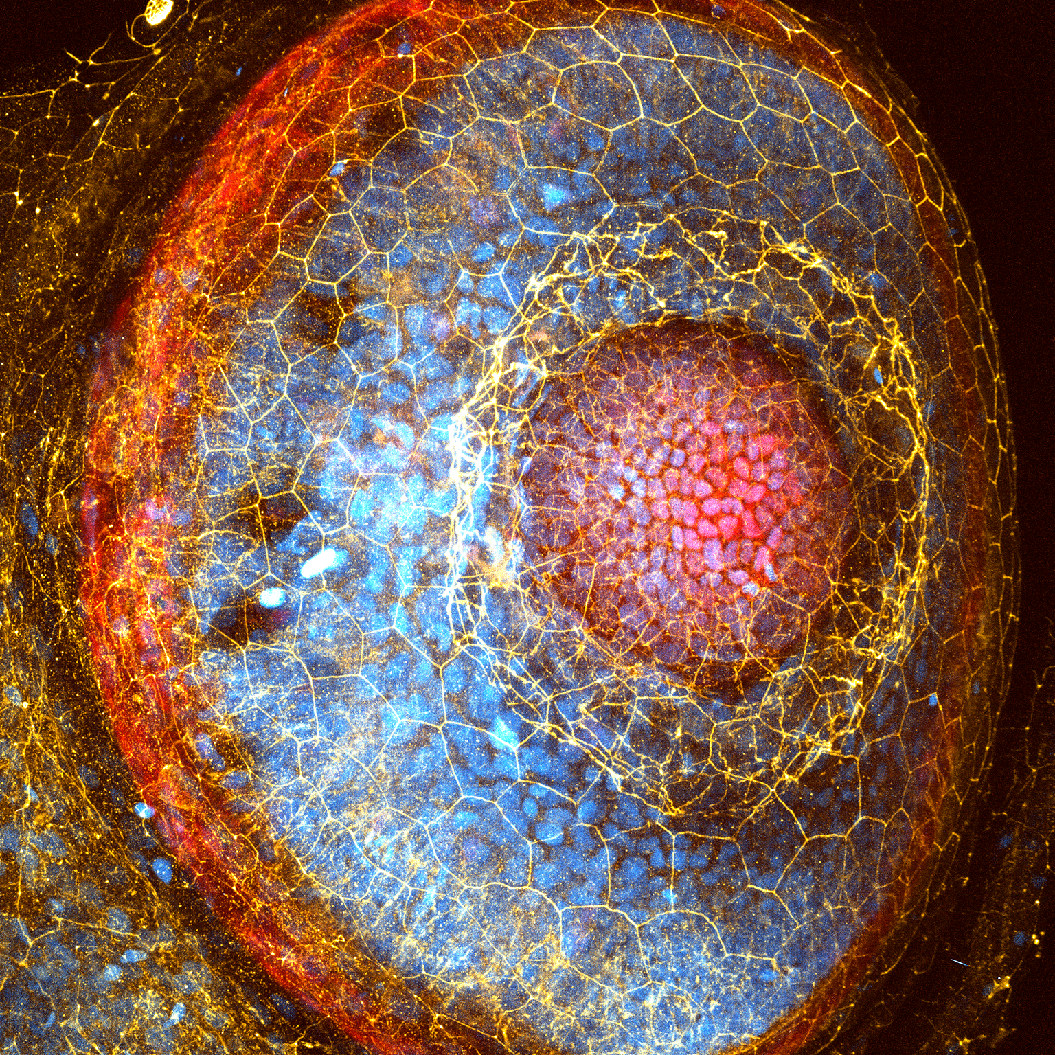
Web of Life
This tiled image was taken with a Zeiss LSM800 confocal microscope. It consists of the mesenteric vasculature from a mutant mouse embryo at E18.5.
The blue staining was CD31 which labels for all blood and lymphatic endothelial vessels. The red staining labels for smooth muscle actin, which helps distinguish between arteries, veins and lymphatic vessels. The yellow staining was PROX1 which labels for lymphatic vasculature and valve structures.
This mutant mouse exhibited abnormal lymphatic vasculature, which were thin-walled, devoid of lymphatic valves and sometimes failed to connect with the mesenteric lymph node at the centre of the "web". It was also noted that this mutation results in perinatal lethality, further emphasising the importance of normal and functioning lymphatic vasculature to sustain life.
Ivan Ngui
Centre for Cancer Biology
University of South Australia

2023 ANZSCDB Image Awards


Fish on Fire
This image depicts the development of lymphatic vascular architecture (grey) and its interplay with a novel transcription factor highly expressed in the fins (red) in a 21 days post fertilization zebrafish. Given the size of the zebrafish at this stage, a special mounting technique was required to ensure survival of the animal during the 4 hour in vivo imaging session. The image itself is composed by 20 single images, captured on Olympus FVMPE-RS Multiphoton, 25x objective. Images were individually processed in Fiji to ensure equal exposure parameters and tiled in Adobe Illustrator.
Andrea Usseglio Gaudi
Peter MacCallum Cancer Centre
& University of Uppsala

Growing Kidney
An E12.5 mouse embryonic kidney expressing Tomato (cyan) in all cell types and GFP (magenta) in nephron progenitor cells was imaged every 5 minutes for 72 hours on a spinning disk confocal. Movies like this provide insight into how progenitor self renewal and differentiation are regulated during organogenesis and inspire new strategies to recreate complex tissues in vitro.
Dr Julie Moreau
Monash Biomedicine Discovery Institute
2022 ANZSCDB Image Awards


Life imitating art
Taking inspiration from the Pop Art movement, this Warhol-esque array of live mouse embryos were captured on a Zeiss LSM780 confocal microscope. Embryos are expressing EB3-dTomato, allowing identification of microtubule growth hotspots as well as Membrane-GFP, showing apical cortical rings which will expand and constrict to compact the embryo during preimplantation development.
Dr Jessica Greaney
Australian Regenerative Medicine Institute, Monash University


Golden orb
The captivating complexity of the microtubule cytoskeleton (yellow) in the early mouse embryo as it intricately organises subcellular components such as lysosomes (purple/cyan), visualised by confocal microscopy (Zeiss, LSM980, Monash Micro Imaging Facility). DAPI (magenta).
Azelle Hawdon
Australian Regenerative Medicine Institute, Monash University
2021 ANZSCDB Image Awards

I Spy With My Little Eye
Ivar Noordstra
Institute for Molecular Bioscience, The University of Queensland
“We use zebrafish to study the biological processes underlying the perception of light. This image highlights the fascinating complexity of the zebrafish eye. It consists of multiple cell types including epithelial cells (red), endothelial cells and neurons. All these cells are tightly linked together through specialized protein complexes like tight junctions (yellow). The combination of junctional and nuclear staining (cyan) beautifully illustrates the incredible diversity of cell shapes.”


Three-dimensional alveolar architecture in an involuting murine mammary gland
Krystyna Gieniec
Single Molecule Science, University of New South Wales
“I am investigating the role of calcium signalling in the function of star-shaped, mammary basal cells as they transition through gestating, lactating and involuting states. Additionally, I am examining the calcium signalling cross-talk between mammary epithelial cells and their immediate cellular environment during gland embryogenesis and tumourigenesis.”
2020 ANZSCDB Image Awards

Vascular abnormalities within the skin of a mouse embryo due to the loss of the cell adhesion molecule, ESAM-1. (Dr Genevieve Secker, CCB, SA)
Dissected flank skin tissue from a fixed E18.5 embryo was blocked in PBS-0.3%/TX100/1% BSA (Block), followed by overnight incubation with primary antibodies (α-Endomucin - Purple, α-Prox1- Yellow and α-Smooth Muscle Actin - Cyan) diluted in block. After extensive washing with PBS-0.3% TX100, the tissue was subjected to overnight incubation with Secondary antibodies (Alexa Fluor® conjugated) diluted in block. After thorough washing, samples were mounted and imaged using a Carl Zeiss LSM 800 Axio Observer 7 confocal microscope. The image was compiled using ZEN 2.5 Blue edition and Adobe Photoshop CC software.

Embryo cells met their fate: to go inside, or stay outside? (Dr Jennifer Zenker, ARMI, VIC)
A plethora of questions are to be answered during life but the very first one, 3 days after fertilisation, is: does a cell go inside or stay outside? Or with other words, become pluripotent or extraembryonic? One cell on the top right of the embryo decided to go inside demonstrated by its apically constricted cell surface (white) while the other cells remained outside. The shapes of the cells are fortified by the microtubule cytoskeleton (golden-red) keeping sister cells connected via a microtubule bridge (golden hot spots at tri-cellular junctions).

Patterning of a wild type mouse tail at the end of axis elongation. (Dr Jan Manent, ARMI, VIC)
The exquisite organization of the developing mouse tail at the end of axis elongation is visualized by light sheet microscopy of a cleared E12.5 mouse embryo stained with Sox2 (neural tube, red), T/Brachyury (notochord, green), and Foxa2 (ingressing tail gut and floor of the neural tube, cyan). Embryos were processed following the 3DISCO protocol (Ertürk, 2012), and imaged on a light sheet Ultramicroscope (LaVision BioTec GmbH). 3D reconstitution, segmentation and animation was performed in Imaris (Bitplane)
Previous Awardees:
2023 - Andrea Usseglio Gaudi, Peter MacCallum Cancer Centre & University of Uppsala | Julie Moreau, Monash Biomedicine Discovery Institute
2022 - Jessica Greaney, ARMI, Monash University | Azelle Hawdon, ARMI, Monash University
2021 - Ivar Noordstra, IMB, University of Queensland | Krystyna Gieniec, University of New South Wales
2020 - Jan Manent, ARMI, Monash University | Genevieve Secker, CCB University of Adelaide | Jennifer Zenker, ARMI, Monash University